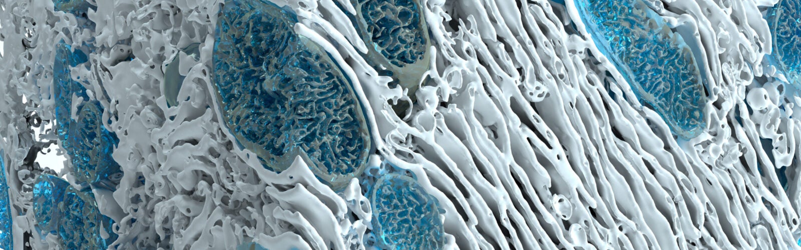
The Sabri Ülker Advanced Imaging Lab possesses instrumentation that was acquired with the generous support of the Ülker family. Their financial support has enabled us to image live and fixed biological samples, such as cell lines and metabolic tissues, under different nutritional conditions – all in high resolution. We are able to capture information such as cell morphology, lipid content, organelle abundance, distribution, localization, motility and interaction. The Sabri Ülker Advanced Imaging Lab contains three systems that allow for the determination of organelle dynamics spatio-temporally by imaging organelles with targeted fluorescent molecules. With these tools we can investigate questions regarding the complex relationship between form and function. The following equipment comprises these three systems:
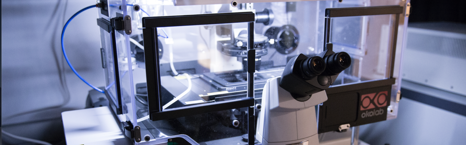
- Spinning disk confocal system: Yokogawa CSU-X1 with a Nikon Ti-E inverted microscope, with a 60x or 100x objective lens equipped with Zyla cMOS camera and iXon EMCCD camera. NIS elements software is available for acquisition parameters, shutters, filter positions and focus control. This system includes a chamber with temperature, humidity and gas controllers to allow live cell and tissue imaging.
- Laser Scanning confocal system: Nikon C2 Laser Scan Confocal with a C2 scan head and controller upgraded high sensitivity, low noise PMT detectors (sensitivity up to 850nm) coupled to a Nikon Ti-E inverted microscope.
- An epifluorescence system coupled to a Ti-S/L100 Inverted Microscope L100 with fluorescent light source (X-Cite 120LED System) equipped with Lambda DG-4 Plus for UV ratiometric probes such as Fura-2 (for calcium measurement) and filters for FRET between ECFP/EYFP
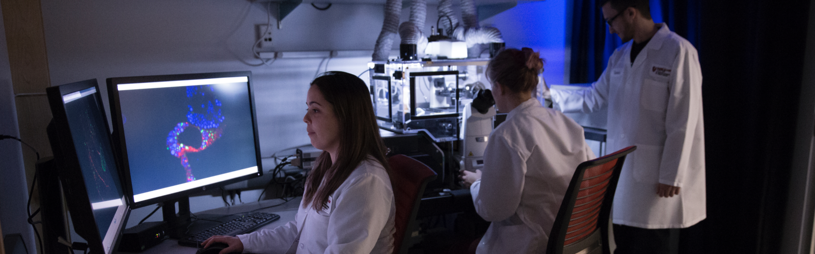
The Advanced Imaging Lab has a high-powered computing work station necessary to compile the massive images produced by these imaging systems. It contains cutting-edge software and hardware to process, analyze and quantify multiple-terabytesize datasets that contain ultrastructural information from electron microscopy and fluorescence microscopy imaging.